Figure 2.

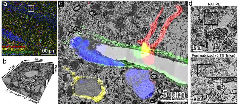
NATIVE enables tissue-scale, multiplex CLEM. (a) Fluorescence confocal image stack is used to identify a region of hippocampus with abundant astrocytes, microglial cells and capillaries, by virtue of nanobody-labeled GFAP (astrocyte marker) in red, CD11b (microglia marker) in yellow, Ly-6C/6G (endothelial cell marker for capillaries) in green and cell nuclei stained by Hoechst in blue. This experiment was performed five times independently, with same results obtained each time. (b) High resolution serial sectioning EM dataset acquired in the region marked by the white box in a. (c) A section in the acquired EM dataset shows that NATIVE-stained tissue is suitable for high quality CLEM. The aligned confocal image layer confirms molecular identities of the different cell types (a fuller demonstration of the alignment between fluorescence and electron microscopy can be seen in Supplementary Video 2. (d) Tissue ultrastructure was compared between NATIVE (top) and full-size antibody staining conditions with 0.1% Triton X100 (bottom). Full resolution pictures are provided in Supplementary Figure 5, Supplementary Figure 6. This experiment was performed twice independently, with same results obtained each time.
