. 2019 Feb 27;8:e43088. doi: 10.7554/eLife.43088

Figure 2. The Atg16 complex interacts with the Atg1 complex.

(A–C, E) Yeast cells expressing Atg5-FLAG (A, C–E) or Atg16-FLAG (B) from each chromosomal locus were treated with rapamycin for 2 hr, and subjected to immunoprecipitation using anti-FLAG antibody. The immunoprecipitates were analyzed by immunoblotting using antibodies against FLAG (A, B), Atg12 (C, E), Atg17 (A–C, E), and Atg1 (C). (D) atg13Δ cells expressing wild-type Atg13, the F375A mutant, or the F430A mutant from centromeric plasmids were treated with rapamycin for 2 hr, subjected to immunoprecipitation using anti-FLAG antibody, and the immunoprecipitates were analyzed by immunoblotting using antibodies against Atg12, Atg13 and Atg17. (F) Yeast cells were treated with or without rapamycin for 2 hr, and coimmunoprecipitation of Atg17 with Atg5-FLAG was examined as described in Figure 2C. (G) Coimmunoprecipitation of Atg17 with Atg5-FLAG was analyzed in cells expressing wild-type Atg1 or the D211A mutant from the original chromosomal locus as described in Figure 2C.

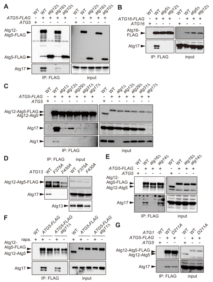
Figure 2.

Figure 2—figure supplement 1. Proteomic analysis to identify proteins bound to the Atg16 complex.

Figure 2—figure supplement 1.

(A) Yeast cells expressing Atg5-FLAG were converted to spheroplasts, treated with rapamycin for 2 hr, solubilized with 1% DDM, and subjected to immunoprecipitation using anti-FLAG antibody. Immunoprecipitates were analyzed by SDS-PAGE, followed by SYPRO Ruby staining. (B) Immunoprecipitates analyzed in (A) were subjected to mass spectrometry, and the MASCOT scores for Atg proteins identified are shown. Proteins with gray background are components of the Atg1 complex. (C) Immunoprecipitates of Atg5-FLAG were prepared as described in (A), and analyzed by immunoblotting using antibodies against Atg12, Atg1, Atg13, Atg17, Atg29 and Atg31.
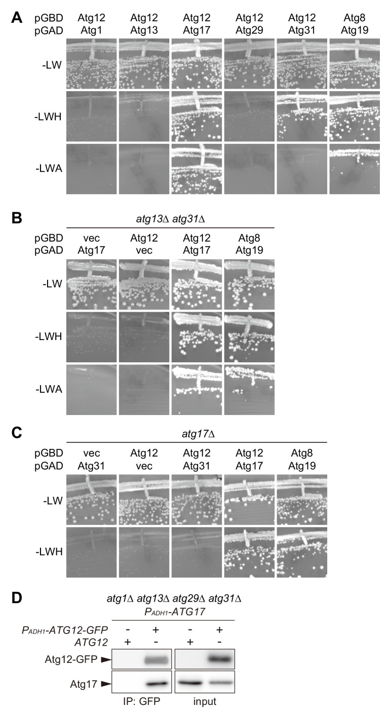
Figure 2—figure supplement 2. Atg17 interacts with Atg12.

Figure 2—figure supplement 2.

(A–C) AH109 cells expressing Atg proteins fused with the Gal4 activation domain (GAD) or Gal4 DNA-binding domain (GBD) as indicated were grown at 30°C for 3 days on SC agar plates lacking leucine and tryptophan for the maintenance of the GAD and GBD plasmids, respectively (-LW), and additionally either histidine (-LWH) or adenine (-LWA) for the assessment of interactions. Cells carrying pGBD-ATG8 and pGAD-ATG19 serve as a positive control. pGAD-C1 and pGBD-C1 were used as vector controls. (D) atg1Δ atg13Δ atg29Δ atg31Δ cells expressing both ATG17 and ATG12-GFP with the ADH1 promoter (PADH1) were treated with rapamycin for 2 hr, subjected to immunoprecipitation using GFP-binding protein-conjugated beads, and the immunoprecipitates were analyzed by immunoblotting using antibodies against Atg12 and Atg17.