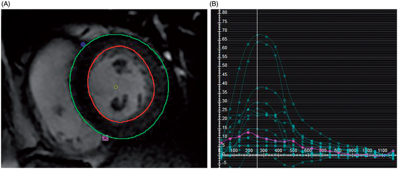
Figure 1.
Radial Strain Calculation MRI Feature Tracking (FT) Strain Measurement. (A) On b-SSFP images, the left ventricular epicardial and endocardial contours are manually traced, with exclusion of the papillary muscles. After tracing multiple short axis levels at diastole, MRI FT software generates multiple regional strain curves shown in (B). The y-axis indicates the unitless strain values and the x-axis measures milliseconds after the R wave. From these regional strains a global radial strain is calculated.

