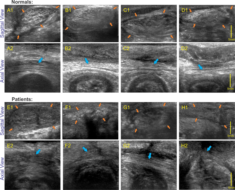
Figure 5:

High frequency sagittal (perineal body) and axial images in normal and patients: upper 2 rows shows sagittal and axial images of the anal canal in 4 normal subjects and bottom two rows show sagittal images in 4 patients with fecal incontinence. Note that in all normal subjects oval shaped perineal body (orange arrows) is clearly visualized with white speckles of different shapes and sizes in axial images and linear white strands (muscle fascicles), marked by blue arrows, in the axial images of the perineal body. On the other hand, perineal body in patients is relatively dark and there is loss and disorganization of linear strands (muscle fascicles).
