Figure 3.
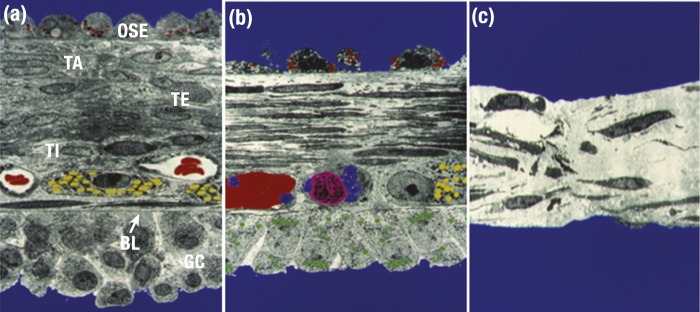
Structure of the apex of the rabbit preovulatory follicle. Faux-colored electron microscopic images were obtained (a) before the LH surge, (b) 1 to 2 h prior to follicle rupture, and (c) immediately before follicular rupture. (a) Layers of the intact follicular wall. At the apex is a single layer of ovarian surface epithelium (OSE) containing granules with unknown contents (red). Underlying the OSE is the tunica albuginea (TA) and theca externa (TE), with numerous cells and extracellular connective tissue. Capillaries with red blood cells (red) and steroidogenic theca interna cells (TI, containing yellow lipid droplets) are adjacent to the granulosa cell (GC) basal lamina (BL). (b) Changes in the follicular wall following an LH stimulus. Notable changes include loss of many of the OSE, elongation of fibroblasts and thinning of the ECM in the TA and TI, and fewer granulosa cells. Capillaries contain clotted red blood cells (red), platelets (blue), and immune cells (pink). Granulosa cells now contain many lipid droplets (green), consistent with increased steroid hormone synthesis. In (c), which depicts the follicular apex immediately prior to ovulation, no OSE or granulosa cells remain at the apex. Remaining connective tissue is thin and disorganized. [Color micrographs courtesy of Dr. Lawrence Espey.]

