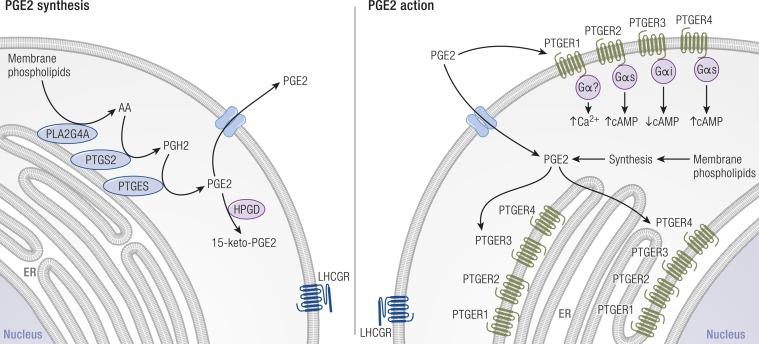
Figure 8.
PGE2 synthesis, receptors, transport, and metabolism in granulosa cells. Left: PGE2 synthesis enzymes (blue) are associated with membranes of the nuclear envelope (not shown) and endoplasmic reticulum (ER). PLA2G4A cleaves arachidonic acid (AA) from membrane phospholipids. PTGS2 converts AA into PGH2. PTGES converts PGH2 into bioactive PGE2. PGE2 is converted to an inactive metabolite (15-keto-PGE2) by HPGD (purple). Right: PGE2 acts via four PGE2 receptors (PTGER1, PTGER2, PTGER3, and PTGER4, green). Each PTGER couples to a subset of G proteins (purple); most frequently used and major intracellular signals are shown for plasma membrane PTGERs. PTGERs can also be located in the membranes of the ER and nucleus; G proteins also couple with PTGERs in these locations (not shown). On both panels, multiple methods of PGE2 transport across the plasma membrane have been proposed (blue) and are discussed in the text. [Adapted with permission from Duffy DM. Novel contraceptive targets to inhibit ovulation: the prostaglandin E2 pathway. Hum Reprod Update 2015;21(5):652–670. Illustration presentation copyright of the Endocrine Society.]

