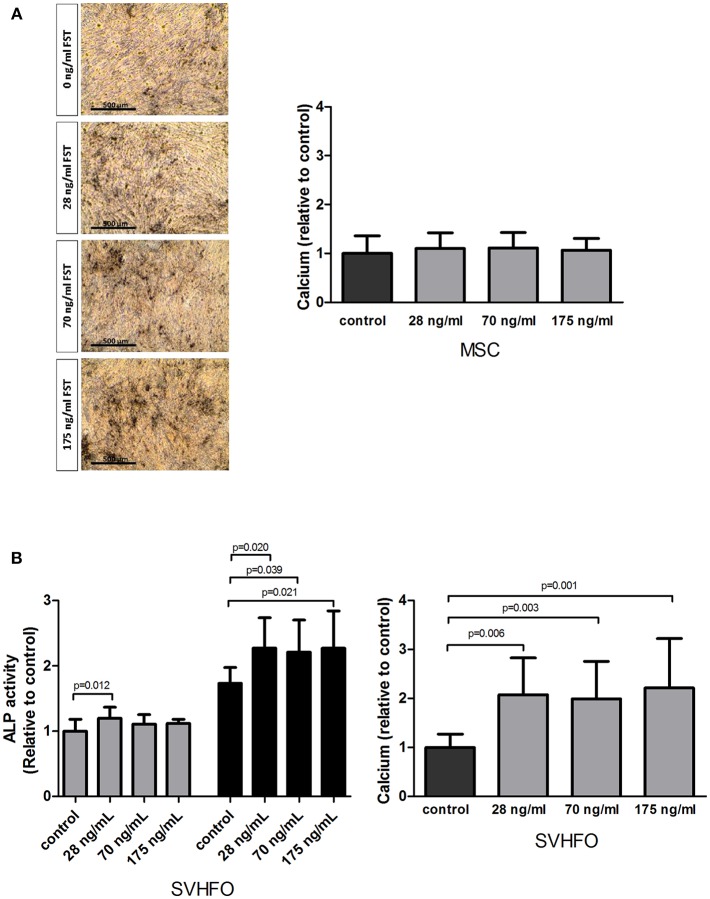
Figure 3.
Effect of follistatin on osteogenic differentiation. Human MSCs and osteoblasts were induced to mineralize in the absence or continuous presence of follistatin (FST315). (A) Quantification of calcium deposition (nmol/ cm2) in the MSC extracellular matrix at the onset of mineralization relative to control (osteogenic differentiation medium) (n = 4 donors performed in triplicate). Donor dependently, mineralization started between 18 and 22 days of culture. Representative pictures of the Von Kossa staining at the onset of mineralization (scale bar: 500 μm). (B) Left graph: alkaline phosphatase (ALP) activity (mU/ cm2) during SV-HFO culture with and without continuous FST treatment at day 9 (gray bars) and 16 (black bars) of culture. Results are shown relative to day 9 control. Right graph: Quantification of calcium deposition (nmol/ cm2) in the SV-HFO extracellular matrix at day 16 relative to control (n = 3 experiments performed in triplicate). The bars show the mean ± SD.

