
Figure 2.
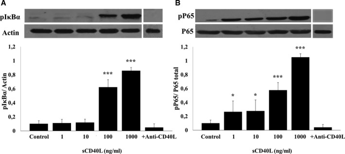
sCD40L phosphorylates IκBα and P65 in human platelets. Washed human platelets (1000×106/mL) were treated with different concentrations of sCD40L (1, 10, 100, and 1000 ng/mL) for 5 minutes at 37°C. Platelets were also pretreated with anti‐CD40L and stimulated with sCD40L (1000 ng/mL). Platelet lysates were resolved in 10% SDS–PAGE and assessed for (A) pIκBα and (B) pP65. Actin blot is from stripped membranes of pIκBα blot; and P65 blot is from stripped membranes of pP65 blot. Blots are representative of 4 independent experiments. Histograms represent the mean of data, expressed in optical density (n=4, mean±SEM). *P<0.05; † P<0.001 (one‐way ANOVA followed by Dunnett's multiple comparisons test vs control).