
Figure 7.
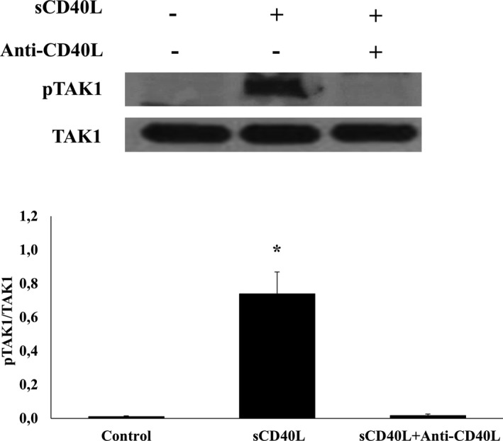
TAK1 is present in platelets and phosphorylated by sCD40L. Washed human platelets (1000×106/mL) were treated with 10 μg/mL of blocking polyclonal anti‐CD40L 37°C, then stimulated with 1000 ng/mL of sCD40L for 5 minutes. Platelet lysates were resolved in 10% SDS–PAGE and assessed for pTAK1. TAK1 blot is from stripped membranes of pTAK1 blot. Blots are representative of 4 independent experiments. Histograms represent the mean of data, expressed in optical density (n=4, mean±SEM). pTAK1 indicates phospho transforming growth factor‐β‐activated kinase 1. *P<0.001 (one‐way ANOVA followed by Dunnett's multiple comparisons test vs control).