
Figure 8.
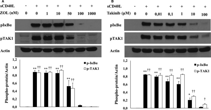
sCD40L activates TAK1/NF‐κB in platelets. Washed human platelets (1000×106/mL) were treated with several concentrations of (A) ZOL or (B) Takinib for 5 minutes at 37°C, and then stimulated with 1000 ng/mL of sCD40L for 5 minutes. Platelet lysates were resolved in 10% SDS–PAGE and assessed for pIκBα and pTAK1. Actin blot is from stripped membranes of pIκBα and pTAK1. Blots are representative of 4 independent experiments. Histograms represent the mean of data, expressed in optical density (n=4, mean±SEM). For pIκBα: **P<0.001, *P<0.01, vs unstimulated platelets. For pTAK1: †† P<0.001, † P<0.01, vs unstimulated platelets (one‐way ANOVA followed by Dunnett's multiple comparisons test vs the unstimulated groups). Zol indicates 5Z‐7‐Oxozeaenol.