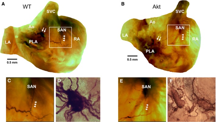
Figure 3.

Acetylcholinesterase staining of wild‐type (WT) and Akita (Akt) atria. A and B, Stained WT and Akita hearts respectively. Pulmonary vein ganglia (PVG) are indicated by white double arrows. Triple arrowheads indicate nerves projecting from PVG to the sinoatrial node (SAN). C and E, Enlarged views of the SAN area boxed in white in WT and Akita hearts. D and F, ×20 magnification of the PVG (white double arrows) in panels A and B. Ao indicates aorta; LA, left atrium; PLA, posterior left atrium; RA, right atrium; SVC, superior vena cava.
