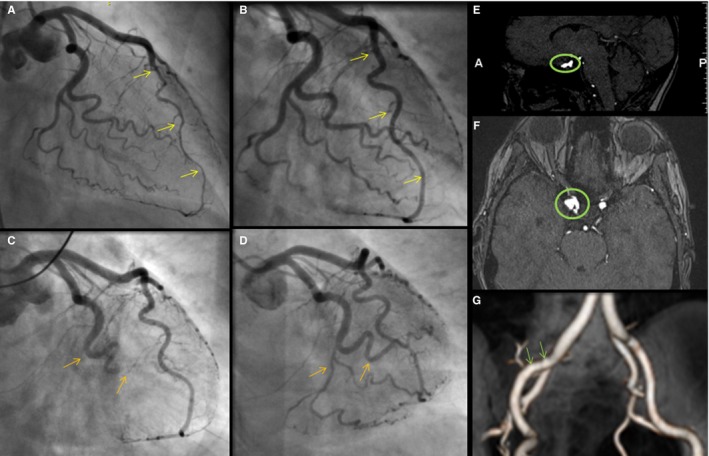
Figure 4.

Imaging of vascular abnormalities and recurrent SCAD in a patient with migraine. This 55‐year‐old female's initial spontaneous coronary artery dissection (SCAD) caused an intramural hematoma of the left anterior descending coronary artery (A, arrows); follow‐up coronary angiography demonstrated interval healing (B, arrows). Several years later, she presented with SCAD of the left circumflex with occlusion of the first obtuse marginal, distal circumflex and its branches (C, arrows). Despite an unsuccessful percutaneous intervention attempt, follow‐up coronary angiography showed interval healing (D, arrows). She also was found to have a 7‐mm right periophthalmic cavernous carotid aneurysm (E and F), 3‐mm left cavernous internal carotid artery aneurysm, 2‐ to 3‐mm right cavernous internal carotid aneurysm, and mild fibromuscular dysplasia of the right external iliac artery (G).
