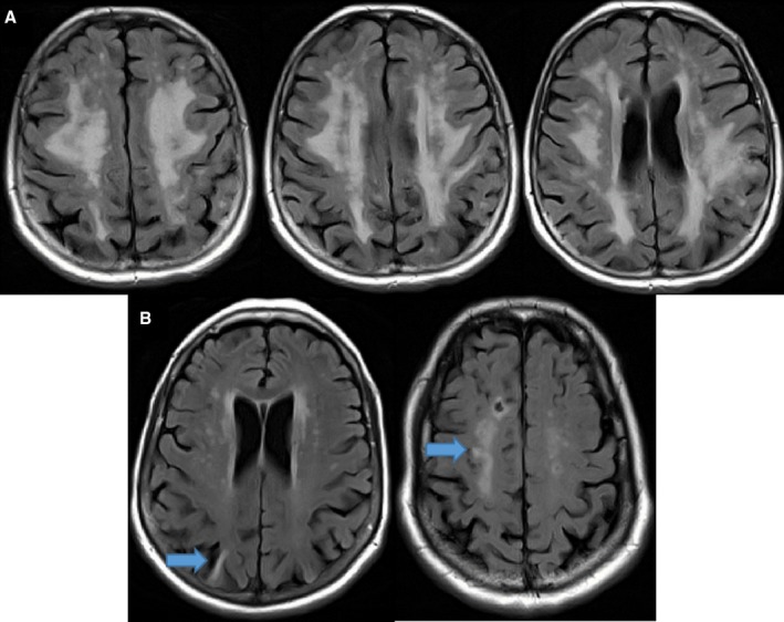
Figure 1.

A, Sample MRI demonstrating white matter hyperintensity (WMH) in a participant. The total calculated WMH volume demonstrated in this axial fluid‐attenuated inversion recovery (FLAIR) is 101.2 cm3, and the total percentage of normal white matter that includes WMH is 19.36%. B, Sample participant's axial FLAIR sequences demonstrating cortical infarction (left arrow) and lacunar infarction (right arrow) as defined by study criteria. MRI indicates magnetic resonance imaging.
