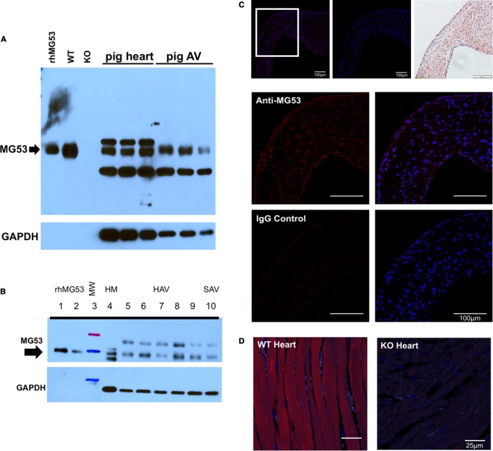
Figure 1.

MG53 is expressed in pig and human patient aortic valve tissue. A, Western blotting shows that MG53 (53 kDa, arrow) is expressed in pig myocardium and aortic valves. rhMG53 (0.03 ng) and wild‐type mouse myocardium (0.06 μg) were used as positive controls and Mg53−/− myocardium (0.06 μg) as a negative control. 10 μg of lysates from pig tissues were loaded from 3 different animals. The apparent lack of GAPDH expression in the mouse myocardial tissue reflects the ≈150‐fold less loading of mouse vs pig protein lysates. B, Western blotting shows that MG53 (53 kDa, arrow) is expressed in human myocardium and aortic valves. rhMG53 was loaded in lanes 1 to 2 (0.05 and 0.02 ng, respectively); ladder in lane 3; non‐failing myocardium (2.5 μg) in lane 4; non‐diseased valves (10 μg) in lanes 5 to 9; and a stenotic valve (10 μg) in lane 10. C, Immunohistochemistry of axial sections of pig aortic valve shows that MG53 (red) is expressed in the valve interstitial layers as well as its endothelial linings. In the top‐most row, ×20 magnification images are shown of (left to right) MG53 staining, rabbit immunoglobulin G control staining, and hematoxylin and eosin staining. In the lower rows and from the ×20 boxed area, ×40 magnification images are shown of MG53 and rabbit immunoglobulin G control staining (red) and overlaid DAPI (4′,6‐diamidino‐2‐phenylindole) staining (blue). D, MG53 staining (red) of wild‐type and Mg53−/− mouse myocardium are positive and negative controls, respectively. AV indicates aortic valve; HAV, healthy aortic valves; HM, healthy myocardium; IgG, immunoglobulin G; KO, knockout (Mg53−/−); MW, molecular weight; SAV, stenotic aortic valve; rh, recombinant human; WT, wild‐type.
