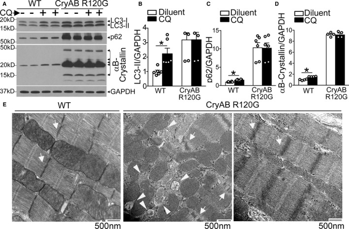
Figure 1.

Autophagic flux is impaired in αB‐crystallin R120G mutant transgenic mice with onset of heart failure. A through D, Representative immunoblots (A) demonstrating expression of LC3 with quantitation of LC3‐II (B), p62 (C) and αB‐crystallin (D) in Myh6‐CryABR120G transgenic mice or littermate wild‐type (WT) controls at 40 weeks of age, injected with chloroquine (40 mg/kg) or diluent to assess autophagic flux. N=4 to 6/group. E, Representative myocardial transmission electron micrographs from in Myh6‐CryABR120G transgenic mice or littermate WT controls at 40 weeks of age. Arrows point to Z‐discs, and arrowheads point to autophagic structures. Representative of N=2/group. *P<0.05.
