Figure 1.

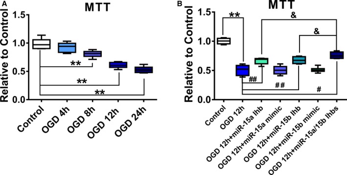
The effect of oxygen and glucose deprivation (OGD) on the proliferation of luc‐mesenchymal stem cells (MSCs) in vitro. A, luc‐MSCs were treated with OGD for 4, 8, 12, or 24 hours. Cell viability was detected by MTT assays. Data are expressed as median (quartile 1–quartile 3), n=6. *P<0.05, **P<0.01 vs Control. B, luc‐MSCs were pretreated with miR‐15a/15b inhibitors or miR‐15a/15b mimics for 24 hours and then cultured under OGD condition for 12 hours. Cell viability was also detected by MTT assays. Data are expressed as median (quartile 1–quartile 3), n=6. **P<0.01 vs Control; # P<0.05, ## P<0.01 vs OGD group; & P<0.05 vs OGD plus miR‐15a/15b inhibitors treated group.
