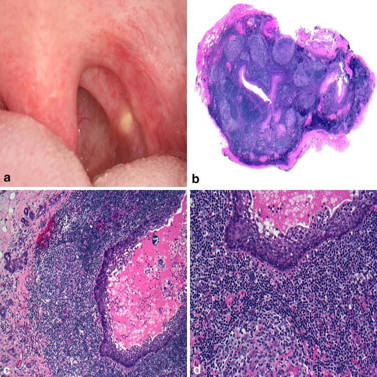
Fig. 2.
Oral lymphoepithelial cyst. a Slightly raised, yellow nodule arising in the tonsillar pillars; b squamous epithelial cyst lining surrounded by hyperplastic lymphoid tissue containing follicular pattern containing germinal centers; c desquamated epithelium and cellular debris filling the lumen; d lymphoid tissue abuts the cystic lining.
Clinical photo courtesy of Dr. Kristin McNamara

