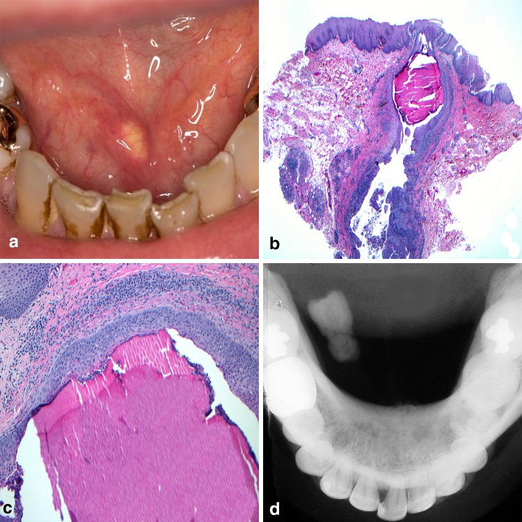
Fig. 5.
Sialolithiasis. a Calcified “stone” within the submandibular gland excretory duct; b sialolith and inflammatory debris within the excretory duct lumen; c calcified mass appearing as concentric deposition; d occlusal radiograph confirms the presence of calcified mass.
Clinical photo courtesy of Dr. Jerry Bouquot

