Fig. 8.
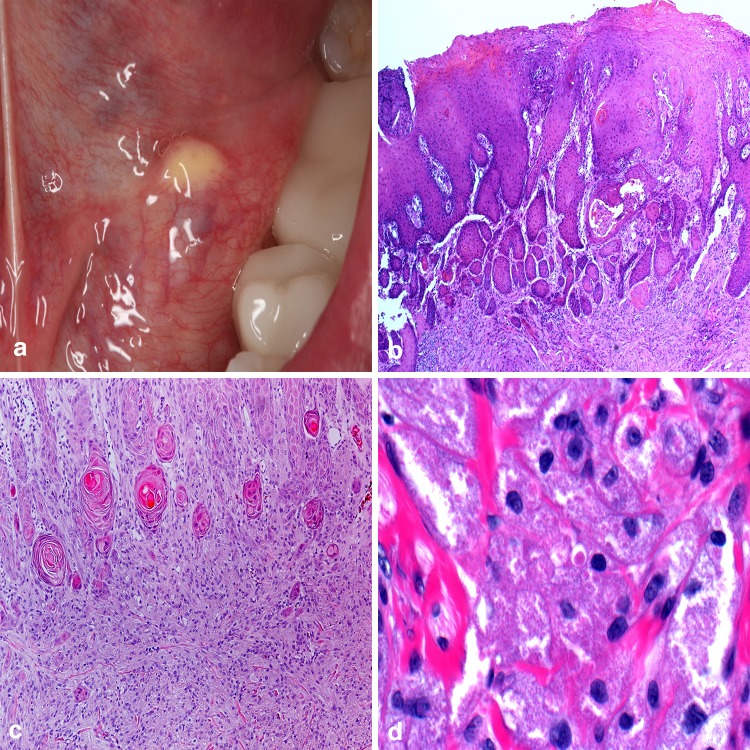
Granular cell tumor. a Firm, yellow dome-shaped mass in the floor of the mouth; b overlying epithelium is nonulcerated and demonstrates pseudoepitheliomatous hyperplasia; c unencapsulated tumor cells arranged in ribbons and sheets around fibrous connective tissue septa and muscle; d large polygonal cells containing numerous autophagolysosomes imparting a course, gritty appearance to the cytoplasm.
Clinical photo courtesy of Dr. Christine Harrington

