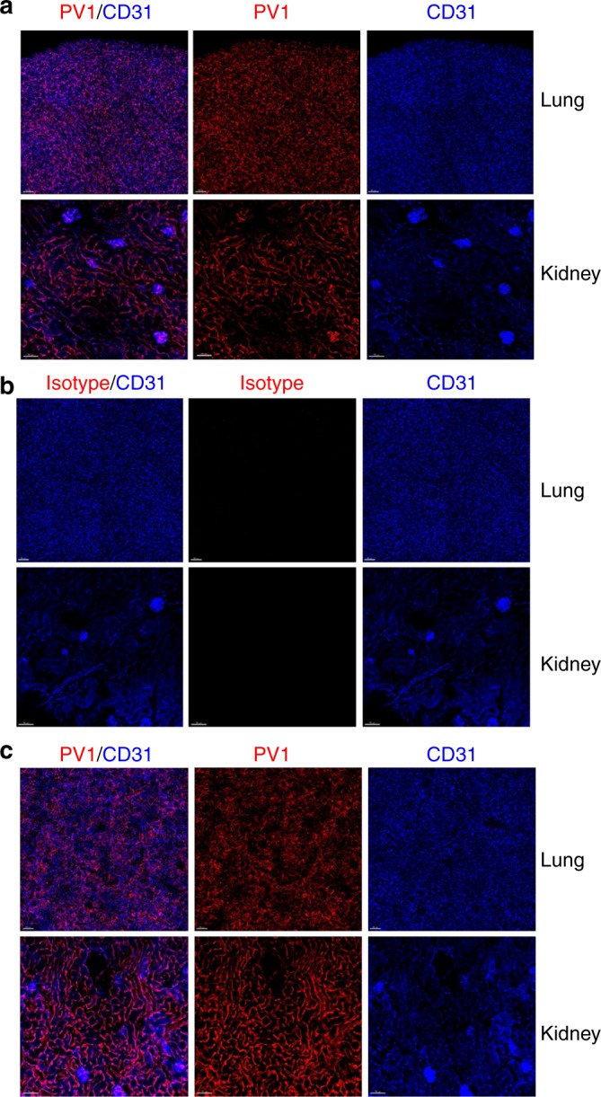
Fig. 3.

Lung and kidney homing of αPV1 antibody captured by confocal microscopy. Mouse lung and kidneys were analyzed by confocal microscopy 4 h after IV administration with either Alexa-594 αPV1 BiS3 (a, c) or Alexa-594 isotype Bis3 (b). Intense PV1 staining (red) can be observed in both untreated (a) and in bleomycin-treated mouse lung and kidney (c). The PV1 staining pattern is similar to the CD31-BV421 staining (blue) pattern revealing a mostly endothelial distribution in the lungs and kidneys. Little to no specific staining in lungs or kidney was observed with an isotype control antibody (b). Images were acquired using Zeiss 880 Airyscan microscope. Bars = 70 µm
