Figure 1.

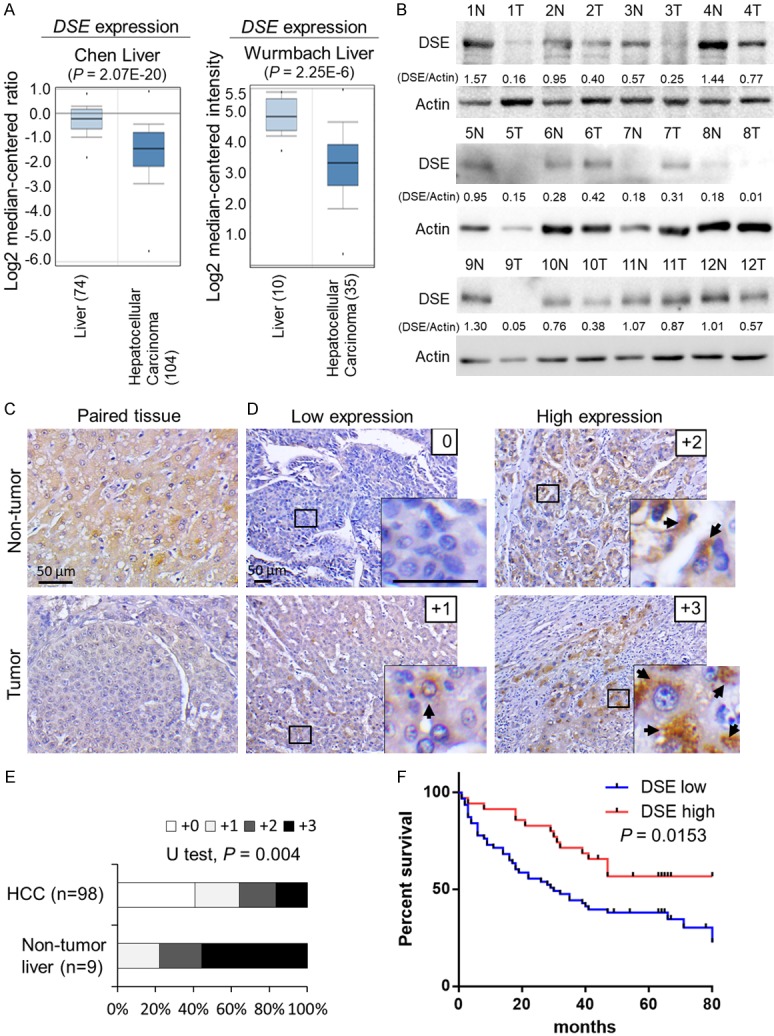
DSE is frequently down-regulated in human HCC and associated with poor overall survival. A. Expression of DSE in the ONCOMINE cancer microarray database. Two independent datasets showed that DSE gene expression is significantly down-regulated in HCC tissue, compared to normal liver tissue. B. Protein expression of DSE in paired HCC tissue. Western blots of DSE using paired non-tumor (N) and HCC tumor tissue (T). Twelve paired samples were tested, and Actin was taken as loading control. Relative quantities are shown. C. Immunohistochemistry of DSE on paired HCC tissue. The staining was visualized in brown color with a 3,3-diaminobenzidine liquid substrate system, and all sections were counterstained with hematoxylin. Representative images of adjacent non-tumor liver (upper) and HCC tumor area (bottom) are shown. Scale bars, 50 μm. D. Intensity of DSE staining on a tissue array comprising 98 primary HCC samples and 9 non-tumor tissue samples. Amplified images are shown at the bottom right. Arrows indicate positive stained HCC cells. Scale bars, 50 μm. E. Statistical analysis of immunohistochemistry in HCC tissue array. Mann-Whitney U Test was used, P = 0.004. F. Kaplan-Meier analysis of overall survival for HCC patients. The analyses were conducted according to the immunohistochemistry of DSE on tissue array. Probability of overall survival was analyzed according supplier’s information. Log-rank test, P = 0.0153.
