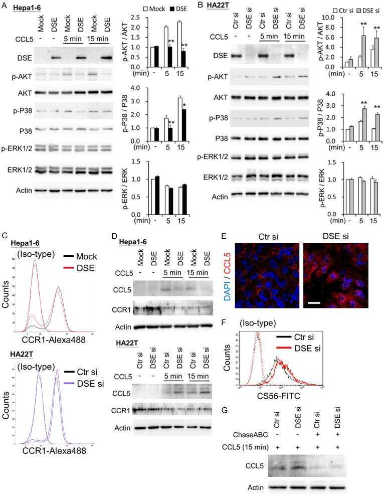
Figure 5.
DSE modulates CCL5-induced signaling and binding in HCC cells. A. Overexpression of DSE decreases CCL5-induced cell signaling in Hepa1-6 cells. B. Knockdown of DSE enhances CCL5-triggered cell signaling in HA22T cells. Cultured cells were serum free starved for 3 hours and then treated with or without (-) CCL5 (50 ng/mL) for indicated time points. Cell lysates (20 μg) were analyzed by western blotting with various antibodies, as indicated. Actin was using as loading control. Representative blots were shown at left. Relative signals were quantified by Image J, and represented as means ± SD from three independent experiments. *P < 0.05; **P < 0.01. C. CCR1 expresses on Hepa1-6 and HA22T cell surface. Transfected cells were stained by CCR1 and anti-rabbit IgG-Alexa488. Non-specific rabbit IgG with anti-rabbit IgG-Alexa488 () was used as iso-type control. D. DSE mediates CCL5 binding on HCC cells. Transfected cells were treated with or without (-) CCL5 (50 ng/mL) for indicated time points. Cells were washed once with PBS and then lysis for western blots with indicated antibodies. Actin was using as loading control. E. Confocal microscopy analysis of CCL5 subcellular localization on HA22T cells. Recombinant CCL5 were treated to control and DSE knockdowned cells for 15 minutes. Cells were stained with CCL5 (red) and nuclei were counter stain with DAPI (blue). Representative images are shown. Scale bars, 20 μm. F. Knockdown of DSE increases CS on HA22T cells. Flow cytometry with anti-CS56 antibody was used. Non-specific mouse IgM was used as iso-type control. G. Enzyme digestion of CS/DS decreases CCL5 binding on HCC cells. Control and DSE knockdowned cells were incubated with or without chondroitinase ABC (ChasABC, 0.5 units/ml) for 3 hours in serum free DMEM. CCL5 (50 ng/mL) were treated for 15 minutes and analyzed by Western blots.

