Figure 4.

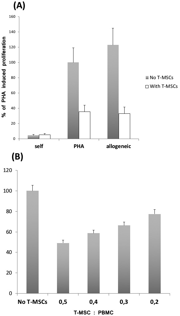
Tonsil-derived mesenchymal stem cells (T-MSCs) inhibited phytohemagglutinin (PHA)-stimulated T cell proliferation in a dose-dependent manner. Peripheral blood mononuclear cells (105 cells) with 5 μg/mL PHA were incubated for 72 hours in the absence or presence of T-MSCs (5 × 10⁴ or varying ratios). (A) Cell proliferation based on [3H]-thymidine uptake. T-MSCs inhibited the T cell proliferation. The value of 100% was set at the proliferative response (counts per minute per culture) of PHA-stimulated T cell proliferation. All values are mean ± standard deviation of triplicates. (B) T-MSCs demonstrated a dose-dependent suppression of PHA-stimulated T cell proliferation. Results (n = 3) are expressed as percentage relative to T cell proliferation without addition of T-MSCs (the value of 100%). Error bars represent standard deviation. Gray bars – without T-MSCs; white bars – with T-MSCs.
