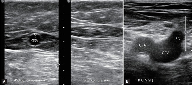
Figure 2.
Sonographic landmark of superficial femoral veins. (A) The ‘Egyptian eye’: a transverse ultrasound image of the great saphenous vein in the thigh with/without compression showing the fascial components that constitute the saphenous compartment. (B) Transverse view of the common femoral vein (CFV) and artery in the right groin: ‘Mickey mouse’ view. GSV, great saphenous vein; CFA, common femoral artery; SFJ, saphenofemoral junction.

