
Figure 5.
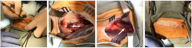
Photography demonstrating a mock surgery on a cadaver sheep for bone chip collection and placement. Left to right: Incision being made toward the distal end of the femur. A 10-mm bone core (arrow) was taken from the distal femur. Bone chips (arrow) were placed on the exposed midshaft of the femur. The incision site was sutured closed. Note that the fascia was also closed by suturing to ensure that the air impact device would not result in surgical site dehiscence.