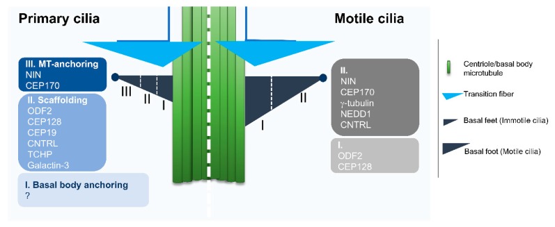
Figure 2.
Comparison of the Structural Organization and major components of basal feet from primary and motile cilia. Primary and motile cilia basal feet present different components and domain organizations as observed by super-resolution microscopy. Primary cilia have multiple basal feet whose components are organized in three distinct domains. The most distal region (III) is characterized by the presence of Ninein (NIN) and Centrosomal protein of 170 kDa (CEP170), proteins involved in microtubule (MT) anchoring and components of the centriole sub-distal appendages. The next region (II) contains proteins that are possibly involved in the scaffolding of the basal feet, namely Outer dense fiber protein 2 (ODF2). The most proximal region (I) to the basal body was named “basal body anchoring” although there was no protein assigned to this region. Motile cilia present only one basal foot, aligned with the cilia beating direction. The domain organization is simpler than that of primary cilia, corresponding, to a certain extent to a rearrangement of Regions II and III. On the most distal region of the basal foot (II), besides the proteins in common with the basal feet of primary cilia (i.e. NIN, CEP170, Centriolin (CNTRL)), there are proteins of the γ-tubulin ring complex (γ-TURC) microtubule nucleating complex (i.e. γ-tubulin and protein NEDD1). The most proximal region (I) contains proteins that are common to the primary cilia basal feet (i.e. Centrosomal protein of 128 kDa (CEP128) and ODF2) that are most likely responsible for the scaffolding (based on proteomics and super-resolution microscopy data in [53]).

