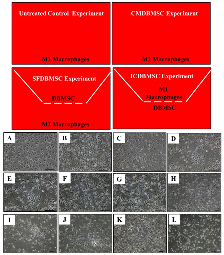
Figure 1.
M1 macrophage culture system. Untreated control experiment consisted of M1 macrophages cultured on a surface of 6-well culture plate in a medium containing GM-CSF, CMDBMSC experiment consisted of M1 macrophages cultured on a surface of 6-well culture plate in a medium containing GM-CSF and CMDBMSC (conditioned medium), SFDBMSC (soluble factor), and ICDBMSC (intercellular direct contact) experiments. In SFDBMSC and ICDBMSC experiment, cells (DBMSCs and monocytes) were separated by transwell chamber membrane culture system. For the experiments of SFDBMSC, DBMSCs were seeded on the upper compartments while monocytes were seeded in the lower compartment. For the experiments of ICDBMSC, DBMSCs were seeded on the reverse side of the membrane while monocytes were seeded on the upper side of the membrane. GM-CSF medium was added to SFDBMSC and ICDBMSC experiments. Effects of human DBMSCs on the morphology of human monocytes differentiated into macrophages by GM-CSF. (A–F) Represent phase-contrast microscopic images showing monocyte (round-shaped morphology) differentiation into M1-like macrophages (fried egg-shaped morphology) after six days of culture in a medium containing GM-CSF (A), in a medium containing GM-CSF and DBMSCs at a 20:1 monocyte: DBMSC ratio (B), at a 10:1 monocyte: DBMSC ratio (C), at a 1:1 monocyte: DBMSC ratio (D), in the presence of 10% CMDBMSC (E), or in the presence of 20% CMDBMSC (F). (G–K) Representative phase-contrast microscopic images showing monocyte-like cells after six days of culture in a medium containing GM-CSF and 30% CMDBMSC (G), 40% CMDBMSC (H), 50% CMDBMSC (I), 60% CMDBMSC (J), 80% CMDBMSC (K), or 100% CMDBMSC (L). Experiments were carried out in duplicate and repeated 30 times using 30 individual preparations of both monocyte-derived macrophages and DBMSCs. Scale bars represent 50 µm.

