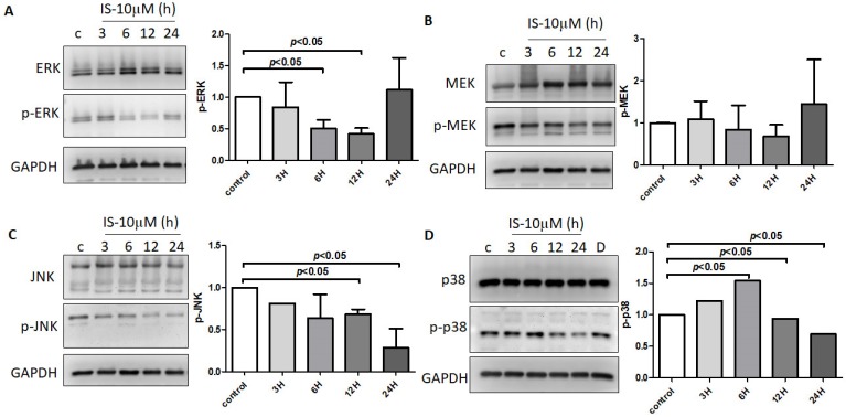
Figure 9.
Human astrocytes treated with control or 10 μM IS for various durations (3 h, 6 h, 12 h, and 24 h). The phosphorylated and total protein levels in the cell lysates were assessed with an immunoblot assay. The results shown are representative of three independent experiments performed on different days, along with relative expression levels to the corresponding control groups at the same time point. IS decreased the phosphorylation of (A) ERK, (B) MEK, (C) JNK, and (D) p-38 at 12 h treatment in human astrocytes. Data represent mean ± SD of a representative experiment.

