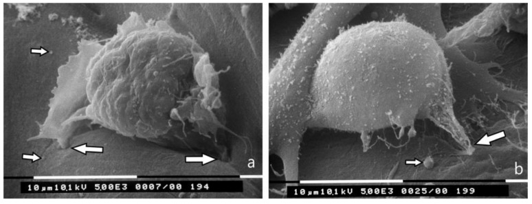
Figure 4.
3D Matrigel (3.0 μg/mL) cell cultures analyzed by SEM. (a) MCF-7 cell lying on a thick layer of Matrigel shows very few microvilli and no microvesicles. Two invadopodia penetrating the Matrigel develop from the ventral side of the cell (large arrows). A few exosomes and microvesicles are shed on the Matrigel surface (small arrows). Bar 10 μm; (b) An MDA-MB-231 cell with few microvilli and microvesicles shows invadopodia (large arrow) penetrating into the thickness of Matrigel. Two adjacent elongated cells with few microvesicles are also visible. Few exosomes and microvesicles (small arrow) shed from MDA-MB-231 cells are present on the Matrigel surface next to the cells. Bar 10 μm.

