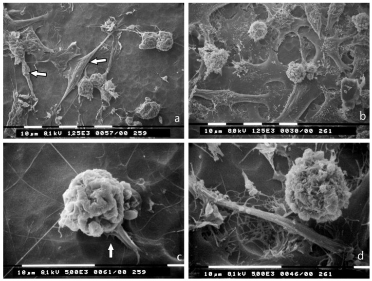
Figure 5.
Breast cancer cells cultured on type I collagen fibrils (lamina propria under basement membrane) observed at SEM. The randomly arranged collagen fibrils completely occlude the holes of the Millipore filter. (a) Several MCF-7 cells showing both globular/spherical and flattened elongated shapes appear isolated or relatively grouped. A few spindle-like cells are also detectable (arrows). Bar 10 μm; (b) MDA-MB-231 breast cancer cells show both globular/spherical and flattened elongated shapes. Bar 10 μm; (c) A globular/spherical MCF-7 cell shows cytoplasmic convolutions, very few microvilli and no microvesicles. Invadosomes from the cell ventral surface are also visible (arrow). Bar 10 μm; (d) A globular/spherical MDA-MB-231 cell in proximity of a Millipore hole covered by sparse collagen fibrils shows microvesicles. Bar 10 μm.

