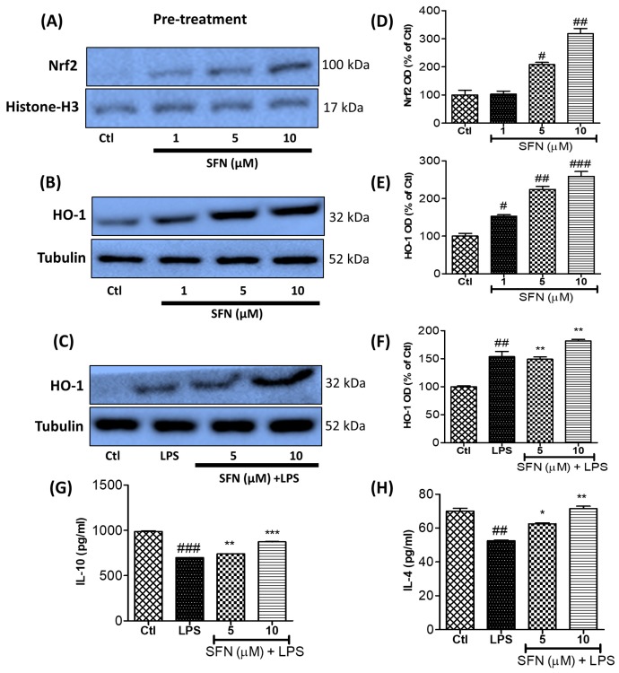
Figure 8.
SFN increased the expression of the anti-inflammatory proteins Nrf2 and HO-1 and the anti-inflammatory cytokines IL-10 and IL-4 in BV2 microglial cells. The cells were pre-treated with SFN for 30 min and then stimulated with LPS (100 ng/mL). After 3 h and 12 h of LPS incubation, the cells were harvested and the expressions of Nrf2 and HO-1, respectively, were determined using western blot analysis. (A–F) Quantification of the Nrf2 and HO-1 expression levels. BV2 cells were pre-treated with SFN, followed by LPS (100 ng/mL) activation for 24 h. The conditioned medium was collected from the treated cells and stored for ELISA evaluation. (G–H) Secreted levels of IL-10 and IL-4, evaluated by competitive ELISA. All data are presented as the mean ± standard error of the mean of three independent experiments. * p < 0.05, ** p < 0.01, and *** p < 0.001 indicate significant differences compared with LPS treatment alone; # p < 0.05, ## p < 0.01, and ### p < 0.001 indicate significant differences compared with the untreated control group. Ctl—untreated control cells; LPS—cells treated with lipopolysaccharide only.

