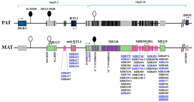
Figure 1.
Localization of miRNA genes with increased expression in male patients with relapsing-remitting multiple sclerosis (in blue color) within DLK1-DIO3 imprinted locus on human chromosome 14q32. The schematic representation of the locus was constructed based on current concept of its structure (according to Human Genome Assembly GRCh38.p12 [22]). Groups of miRNA genes located in close proximity to each other or referred to one structural unit of genome are pooled in lists. Altogether, this locus harbors two large clusters of miRNA genes (10 miRNA genes in 14q32.2 and 44 miRNA genes in 14q32.31). Underlined miRNAs were further taken for validation analysis. The DLK1-DIO3 imprinted locus contains three paternally-expressed protein-coding genes: DLK1 (Delta-like homolog 1), which is located at the 5′-end and encodes for a protein involved in the Notch signaling pathway [23], DIO3 (type III iodothyronine deiodinase) located at the 3′-end and involved in controlling thyroid hormone homeostasis [24] and RTL1 (Retrotransposon Gag Like 1). Fully complementary antisense transcript anti-RTL1 is expressed from the maternal chromosome and acts as a repressor for RTL1. The locus also contains long non-coding RNAs genes (MEG3, MEG8, MIR381HG, MEG9), coming from the maternal chromosome. MEG8 harbors a tandemly repeated array of the C/D-box snoRNA family, namely SNORD112, SNORD113, SNORD114, consisting of one, nine and 31 paralogous copies, respectively. The expression of genes is regulated via imprinting control regions which harbor differentially methylated regions (DMRs): the intergenic DMR (IG-DMR), MEG3-DMR and MEG8-DMR. The IG- and MEG3-DMRs are methylated on the paternal and unmethylated on the maternal allele, while the MEG8-DMR is oppositely methylated on the maternal allele [25]. PAT and MAT stand for paternal and maternal chromosomes; DMR—differentially methylated region. Filled ellipses represent methylated DMRs, and unfilled represent unmethylated DMRs. Blue rectangles represent protein-coding paternally-expressed genes, and green—maternally-expressed non-coding genes; gray rectangles represent repressed genes. Black strips indicate for paternal repressed miRNA and SNORD genes; pink and violet strips indicate maternally-expressed miRNA and SNORD genes, respectively.

