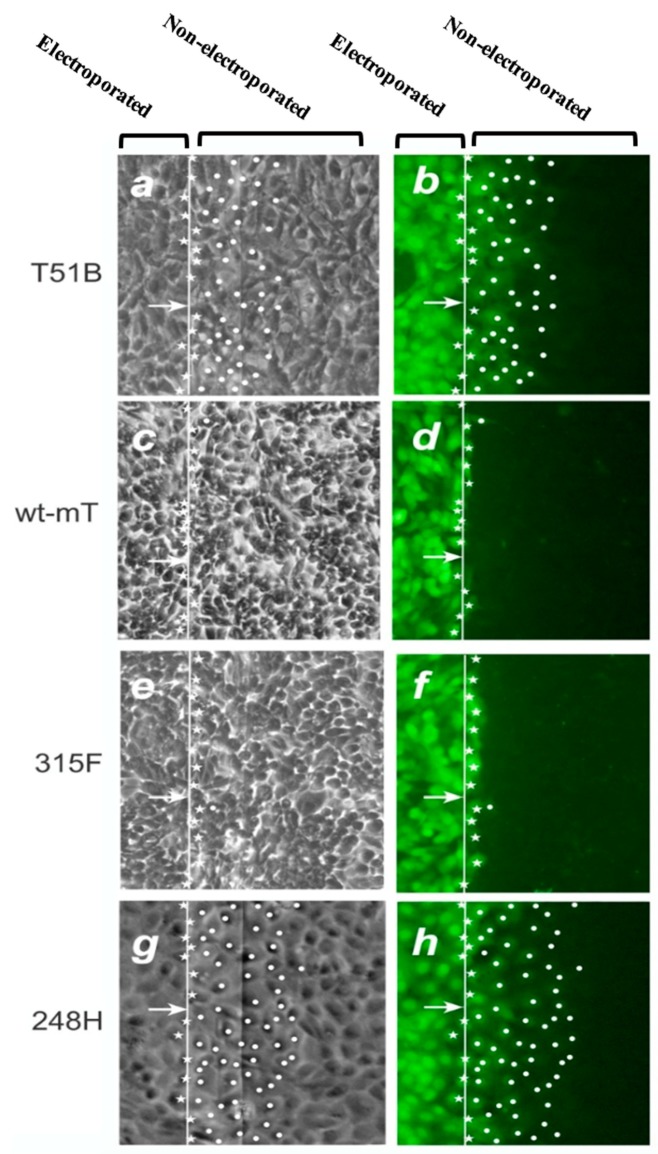
Figure 1.
GJIC in T51B rat liver epithelial cells and derivatives expressing wt or mutant mT. GJIC was examined using a technique of in situ electroporation: The indicated cell lines were grown on a glass slide, part of which is coated with a thin layer (~800 Å) of electrically conductive and transparent Indium-Tin oxide, as shown at the top. The fluorescent dye, Lucifer yellow (LY) was added to the cells and introduced via an electric pulse which causes the formation of “pores” on the plasma membrane. The migration of LY to the neighboring, non-electroporated cells through the cells’ gap junctions is observed under fluorescence illumination (b,d,f,h) and offers a quantitative measure of GJIC (reviewed in [59]). (a,c,e,g): Phase-contrast images of the same fields. Stars indicate cells loaded with LY by electroporation. Dots denote cells where LY has penetrated through gap junctions. Arrows point to the edge of the electroporated area. Magnification: 240x. Note the extensive GJIC of T51B-248H cells. (From [31], reproduced with permission).

