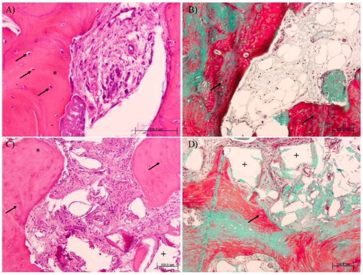
Figure 3.
Histomorphometric analysis of samples from PRF-L (A,B) or β-TCP (C,D) treated sockets (hematoxylin and eosin (A,C) and Masson trichromic (B,D) stainings). Note that more osteocytes in the newly formed bone (arrows) and more new mineralized tissue (*) are present in the PRF-L group. Remnant particles (area left after demineralization, +) are only observed in the β-TCP group.

