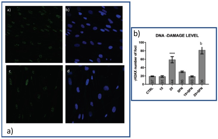
Figure 3.
Effects of SFN alone or in combination with hydrogen peroxide on DNA-damage Subconfluent cells were treated as described in Figure S1 and Material and Methods Section. (a) An example of immunofluorescence obtained with cells untreated (CTRL) and treated with 25 M hydrogen peroxide in presence or absence of SFN. Scale bar is 10 M. Untreated cells (a,b); and treated ones (c,d) stained for H2AX (1:700, Abcam, ab2893-Phospho139) (a,c) or both for H2AX and DAPI (b,d). Cells were fixed with 3.7% paraformaldehyde, permeabilized with 0.1%TRITOX-100 in PBS for 15 min at RT and incubated overnight at 4 C with the H2AX (1:700, Abcam, ab2893-Phospho139). The samples were then incubated with FITC anti-Rabbit (1:250, ab150077, AbCam) for 1 h at RT and mounted with Pro-long anti-fade reagent (P7481, Life Technologies) with DAPI to stain the nuclei. The images were acquired with a Leica TCS NT confocal microscope. Panel b shows H2AX spots inside the nuclei counted using spot detector tool of ICY Software as described in the Materials and Method section. All the resulting values are normalized with the total number of pixels of their image, to make possible the comparison of all the nuclei, one with each other. In the bars are reported the number of cells analyzed for each conditions. **** p < 0.0001 versus untreated cells; b: p < 0.0001 versus SFN treated cells.

