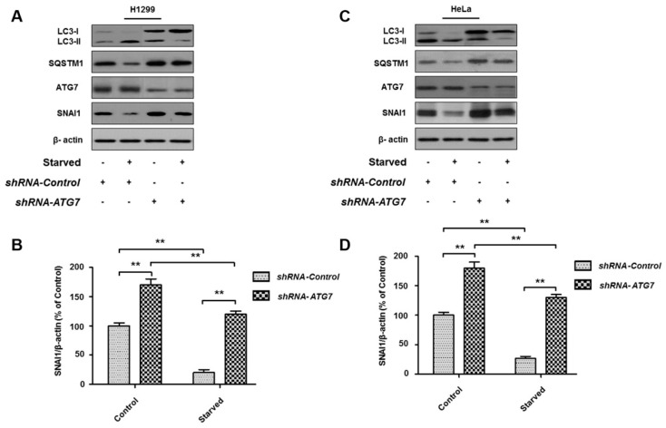
Figure 4.
ATG7 knockdown rescues SNAI1 degradation. H1299 (A) or HeLa (C) cells were transiently transfected with ATG7 shRNA and control shRNA plasmids using Lipofectamine 3000 and incubated for 24 h. Cells were then starved with HBSS for 4 h. Total-cell proteins (30 µg) were separated by 10% or 12% SDS-PAGE and analyzed by western blotting using antibodies against LC3, SQSTM1, ATG7, and SNAI1. β-actin was used as a loading control. (B,D) Quantification of SNAI1. The levels of SNAI1 in H1299 cells (B) and HeLa cells (D) were quantified using NIH ImageJ software. β-actin was used as a loading control. Data represent the mean (±S.D.) of three independent experiments (** p < 0.01).

