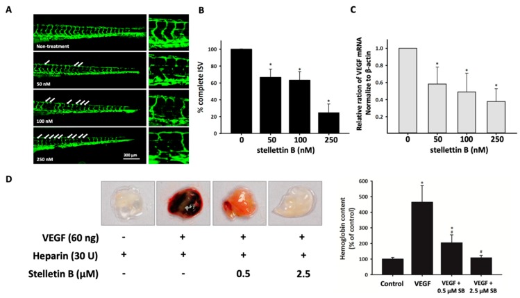
Figure 7.
Stellettin B inhibits angiogenesis in zebrafish and mouse models. (A) Lateral view of Tg (fli1:EGFP)y1 zebrafish embryos at 72 hpf immersed in Hank’s buffer with 0, 10, 50, 100, or 250 nM stellettin B treatment. Scale bar = 300 µm. (B) Percentage of complete intersegmental vessels (ISVs) in each group. Values are expressed as the mean ± SD (n = 12). (C) Real-time quantitative polymerase chain reaction (PCR) analysis of VEGF expression. Values are expressed as the mean ± SD (n = 3). * p < 0.05 relative to the nontreatment group. (D) Effect of stellettin B on in vivo angiogenesis, determined using the Matrigel plug assay. Here, 400 µL Matrigel containing 30 U heparin with/without VEGF and/or stellettin B was subcutaneously injected into BALB/c mice. After 10 days, the Matrigel plugs were removed and photographed. The neovascularization effect on the Matrigel was evaluated through measurement of the hemoglobin content. The results are reported as the mean ± SD (n = 4). * p < 0.05 relative to the control group and # p < 0.05 relative to the VEGF-only group.

