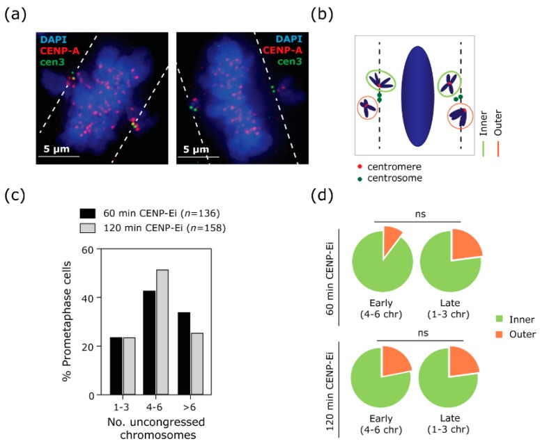
Figure 2.
The position of uncongressed chromosomes (chr) does not fully explain their persistence near the centrosomes. (a) Example immunofluorescence image of RPE1 cells treated for 2 h with CENP-Ei, stained with CENP-A antibody to mark centromeres (red) and with Centrin3 (cen3) antibody to mark centrosomes (green). In the example, two prometaphases are reported: the left panel shows a prometaphase cell in an early stage (4–6 uncongressed chromosomes), the right panel shows a prometaphase cell in a later stage (1–3 uncongressed chromosomes). A white dashed line, parallel to the metaphase plate, has been used as a reference to categorise the uncongressed chromosomes (also schematised in (b)). (b) Any centromeric signal that is touching the line or is located between the line and the metaphase plate is classed as “inner”; any centromeric signal which is not touching the line or is distal from the cen3 signal is classed as “outer”. (c) Distribution of prometaphase cells with 1–3, 4–6 or more than 6 uncongressed chromosomes at each time point (60 min and 120 min) (n = number of cells analysed in each condition). (d) Quantification of position of uncongressed chromosomes in early or late phase cells at 60 or 120 min of CENP-Ei treatment, two experiments (125 cells and 676 chromosomes analysed for 60 min treatment; 126 cells and 717 chromosomes analysed for 120 min treatment). t-test on early vs late cells treated for 60 min, p = 0.3065. t-test on early vs late cells treated for 120 min, p = 0.8987. ns: not significant.

