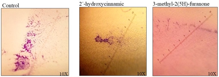
Figure 7.
The appearance of the biofilm of K. pneumoniae in a polyvinyl chloride (PVC) urethral catheter. Biofilm formed at 30 h of incubation at 37 °C was stained with violet crystal at 0.05%. The size of microcolonies was determined in three fields to quantify the colonization level; each image is a catheter field with greater biofilm formation. A 60.15% reduction in colonization was observed in the presence of 2´-hydroxycinnamic acid, and a 67.62% reduction with 3-methyl-2(5H)-furanone at 15 µg/mL was observed.

