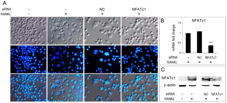
Figure 1.
Inhibition of osteoclastogenesis by silencing of NFATc1. Untransfected, siRNA-non correlated (NC) and siRNA-NFATc1 transfected cells were cultured with RANKL (50 ng/mL) for 24 h. Control untransfected cells were cultured without RANKL. (A) Cells were fixed, stained with DAPI (which stains the nuclei blue) and observed by DIC (upper row) and immunofluorescence (middle row) microscopy. Bottom row shows merged images. (B) Quantitative PCR (QPCR) of NFATc1. mRNA expression was presented as relative values to those expressed in untransfected cells (−/+RANKL) arbitrarily set at 1.0. The results shown are the means ± SD of two experiments (each of which was performed in triplicate). *** p < 0.005. (C) Western blot of NFATc1 protein in untransfected (−/− RANKL) and (−/+ RANKL), siRNA-NC and siRNA-NFATc1 transfected cells (+RANKL). The data shown represent two independent experiments with comparable outcomes.

