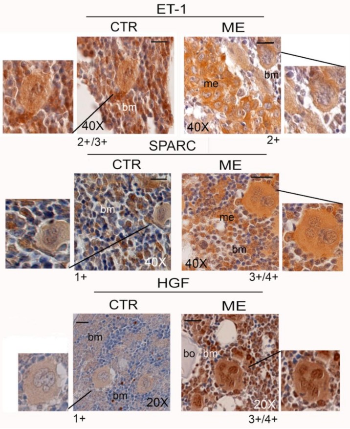
Figure 3.
Positive reaction of MKs for endothelin-1 (ET-1), SPARC, and hepatocyte growth factor (HGF) in the bone marrow of femurs of control (CTR) and bone metastasis-bearing mice (ME). Representative images are shown, and three serial sections were analysed (n = 3). Semi-quantitative analysis of MK immunostaining is given below each panel: 4+ denotes very strong staining; 3+ strong staining; 2+ moderate staining; and 1+ weak staining. Statistical analysis is reported in supplementary materials (Table S1B). me: bone metastasis; bm: bone marrow; bo: bone. Scale bar = 20 μm.

