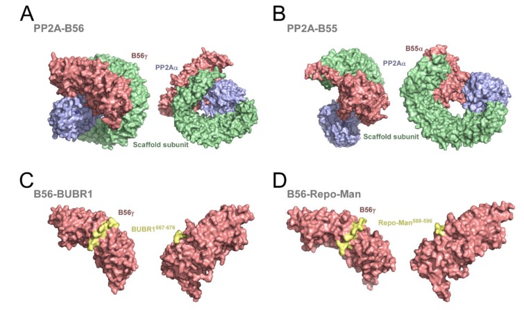
Figure 2.
Structure of PP2Aα holoenzymes. (A,B) Surface representation of the three-dimensional structure of the PP2Aα catalytic subunit (light blue) in complex with the scaffold subunit and the regulatory subunits, B56γ (A) or B55α (B). The PP2A catalytic subunit is represented in light blue with manganese atoms in the active site represented as dark blue spheres. The scaffold subunit is represented in green and the regulatory subunits are depicted in light red. Protein Data Bank (PDB) accession ID 2NPP (A) and 3DW8 (B). (C,D) Surface representation of the three-dimensional structure of B56γ (light red) interacting with BUBR1667–676 and Repo-Man588–596 peptides (yellow). Both peptides encompass a LxxIxE motif that binds to a conserved basic pocket at the concave surface of B56γ. PDB accession ID 5K6S (C) and 5SW9 (D).

