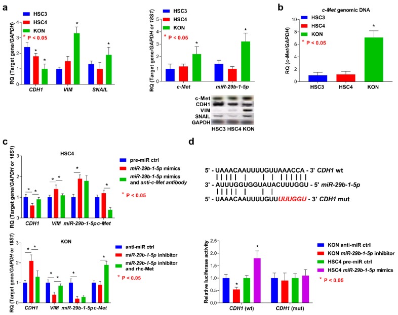
Figure 3.
Analysis of the status of the EMT and c-Met and miR-29b-1-5p expression and function of miR-29b-1-5p in OSCC cells. (a) The levels of expression of miR-29b-1-5p and c-Met in KON cells were higher than those of HSC3 and HSC4 cells (left). Upregulation or downregulation of CDH1 or SNAI1 and VIM was detected in KON cells, respectively (right). (b) MET genomic DNA was amplified compared with the copy numbers measured in HSC3 and HSC4 cells. (c) Induction of the EMT was mediated through the interaction of miR-29b-1-5p with c-Met in OSCC cells. (d) The 3′-UTR of CDH1 contains miR-29b-1-5p binding sites. Reporter constructs showing wild-type (wt) and mutated (mut) CDH1 3′ UTR sequences (top). Luciferase activity of OSCC cells transfected with reporter vectors containing the wt or mut 3′-UTR of CDH1. Error bar: standard deviation (bottom). RQ: relative quantitation.

