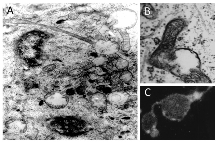
Figure 8.
Local mitochondrial swellings in situ. (A) Electron microscopic picture of a cell from a kangaroo rat kidney epithelium exposed to high hydrostatic pressure (100 mPa). Note multiple local mitochondrial expansions (local mitochondrial swellings) over a single mitochondrial filament with continuous matrix; electron microscopic and fluorescent microscopic evidence (B,C), respectively of a local swelling of mitochondrial filaments in pig embryo kidney epithelial cells after exposure to diazepam. Rhodamine 123 staining in C; note that mitochondrial stain is localized in spots adjacent to the membrane. Used methods as described in [54].

