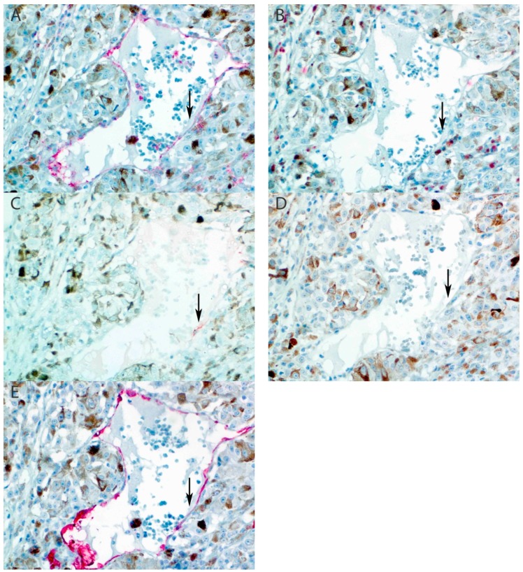
Figure 2.
Sample 15 of Table 2: intratumoral focal positive staining of tumor vasculature for LYVE-1. (A) CD31 stains all endothelial cells (arrow). (B) D2-40 is negative in endothelium. (C) LYVE-1 shows focal positive staining in a large tumor vessel. (D) Prox-1 is negative. (E) CD34 stains all endothelial cells. (All panels: original magnification 400×).

