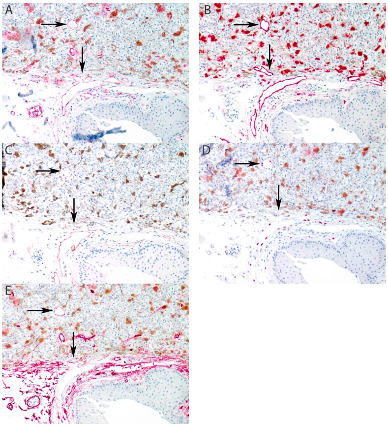
Figure 3.
Recruitment of lymphatic vessels into extraocular extension of uveal melanoma (arrows). (A) CD31 stains all endothelial cells. (B) D2-40 stains conjunctival lymphatic vessel endothelium and demonstrates intratumoral recruitment. (C) LYVE-1 stains conjunctival lymphatic vessel endothelium and demonstrates intratumoral recruitment. (D) Prox-1 is positive in the nuclei of lymphatic endothelial cells and demonstrates intratumoral recruitment. (E) CD34 is positive in blood vessel endothelium and negative in lymphatic endothelium. Note that the intratumoral recruited lymphatic vessel stains weakly positive at the recruitment front (vertical arrow) and intatumoral (horizontal arrow). (All panels: original magnification 100×).

