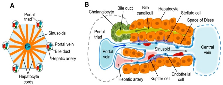
Figure 2.
Liver structure and cell types (A) Functional unit of the liver lobules; Each lobule is composed of a central vein (CV), the portal triad consists of a portal vein, biliary duct and hepatic artery. Hepatocyte cords are separated by sinusoids that carry blood from the portal triads to the central vein. (B) Hepatocytes secrete bile salts into the bile canaliculi that lead to the bile duct. Stellate cells are in the space of Disse between the hepatocyte cords and sinusoids. Kupffer cells, which are the specialised macrophages of the liver, are also located in sinusoids. Epithelial cells lining the bile ducts are called Cholangiocytes (Adapted from Gordillo et al. [6]).

