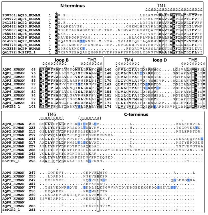
Figure 3.
Sequence alignment of human AQP0-AQP9 and spinach aquaporin SoPIP2;1. The corresponding UniProt IDs are marked in the top panel. Secondary structure information was based on sheep AQP0 (PDB code 2B6O). The alignment was generated using PSI/TM-Coffee server [92] and visualized using ESPript 3.0 [93]. Residues that are strictly conserved are highlighted with black background and similar residues are highlighted in boxes. Phosphorylation sites are highlighted in blue.

