Figure 4.
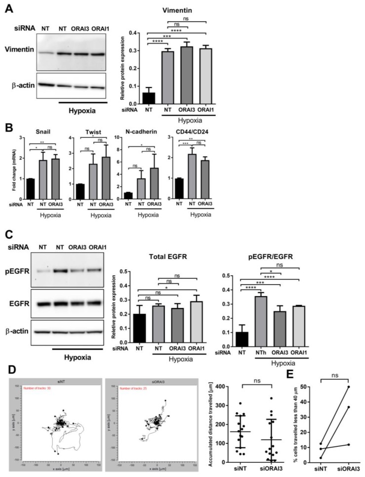
Effect of ORAI3 silencing on the levels of hypoxia-induced expression of EMT markers, EGFR activation and cell migration of MDA-MB-468 breast cancer cells. (A) Representative immunoblot (left) and densitometry analysis (right) of the effect of ORAI3 and ORAI1 silencing on hypoxia-induced vimentin protein expression and (B) the effect of ORAI3 silencing on the mRNA expression of key hypoxia-induced EMT markers in MDA-MB-468 breast cancer cells. (C) Representative immunoblot (left) and densitometry analysis (right) of the effect of ORAI3 and ORAI1 silencing on the total levels of EGFR protein and hypoxia-induced EGFR phosphorylation. ns = not significant (p ≥ 0.05), * p < 0.05, ** p < 0.01, *** p < 0.001, **** p < 0.0001, (one-way ANOVA, with Tukey’s multiple comparisons), n = 3, mean ± SD. (D) Representative spatial plot of all NT control- and ORAI3-silenced cells from one well of the same experiment (left) and quantitative analysis of the distance travelled by 15 randomly selected cells from three independent experiments (5 cells from each experiment). (E) Quantitative analysis of the percent of cells in each experiment from three independent experiments that travelled less than 40 µm. Total number of cells from three independent experiments analyzed were 95 and 80 cells for siNT and siORAI3, respectively. Cell migration was analysed in hypoxic conditions over a period of 12 h after exposing cells to 72 h hypoxia. ns = not significant (p ≥ 0.05) (Mann-Whitney U-test), n = 3, mean ± SD.

