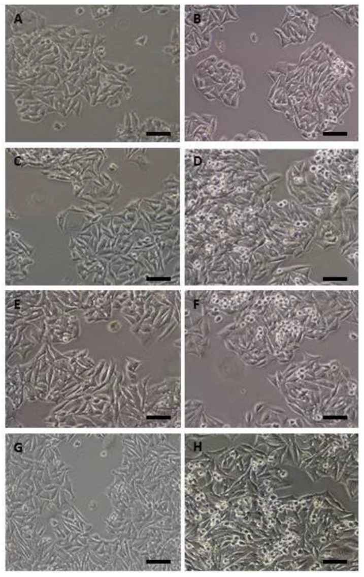
Figure 2.
Phase contrast micrographs of parental DLD-1, DLD1/ Tet-On-gef, DLD1/ Tet-On-apoptin, and DLD1/ Tet-On-gef-apoptin cells after 48h of treatment with doxycycline (Scale bar = 100 μm). No differences were observed between DLD1cells (A) and cells treated with Dox (B). However, the lines transduced with gef and/or apoptin and induced with Dox were characterized by the presence of a group of smaller, rounded cells (more evident in D, F, H), less adhered to the culture bottle when compared to the parental cells. (C) DLD1/Tet-On-apoptin not induced with Dox; (D) DLD1/Tet-On-apoptin induced with Dox; (E) DLD1/Tet-On-gef not induced with Dox; (F) Tet-On-gef induced with Dox; (G) DLD1/Tet-On-gef-apoptin not induced with Dox; (H) DLD1/Tet-On-gef-apoptin induced with Dox.

